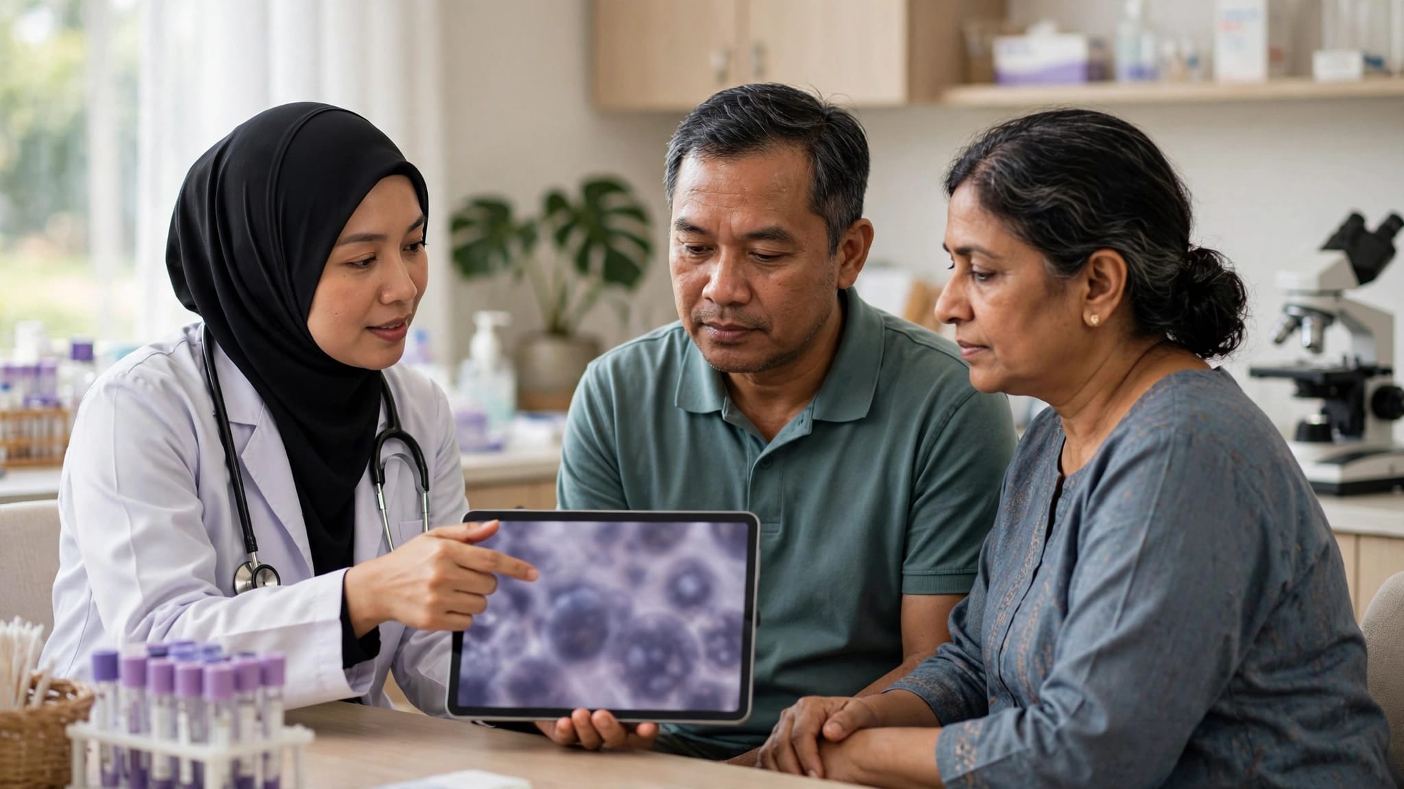
Waiting for results from a skin biopsy can make a few medical terms feel overwhelming. Melanoma biopsy results do more than confirm whether a suspicious mole is cancerous. They help your doctor understand how deeply melanoma cells have entered the skin and guide discussions about staging and next steps.
For patients and families in Malaysia, the most useful first step is to get a copy of the pathology report and discuss it with the doctor who arranged the biopsy. Start with the diagnosis, Breslow thickness, ulceration, and margin status. These findings guide discussion, but they don’t determine an individual prognosis on their own.
Quick Answer
A pathology report for suspected melanoma skin cancer confirms whether the sampled skin lesion is benign, melanoma in situ, or invasive melanoma. For invasive melanoma, breslow thickness and ulceration are important details for cancer staging and treatment planning.
Breslow thickness measures the depth of melanoma in millimetres. Ulceration is a microscopic finding showing that the skin surface over the melanoma has broken down. These findings, along with assessment of lymph nodes and scans where needed, help your specialist decide whether wider surgery or further tests may be appropriate.
5 Key Takeaways
- A positive biopsy result identifies melanoma cells in the sampled tissue. It doesn’t, by itself, show whether cancer has spread elsewhere.
- Tumour depth is measured in millimetres and is a major factor in assessing localised melanoma.
- The microscopic surface finding isn’t the same as a biopsy wound or a visible scab. It is a specific feature identified under the microscope.
- Involved surgical margins mean melanoma cells reach the edge of the removed sample. A wider local excision may be needed, but this doesn’t prove the cancer has spread.
- A sentinel node biopsy isn’t needed for every melanoma. Your doctor considers tumour depth, the microscopic surface finding, your health, and the full pathology report.
Understanding Melanoma Biopsy Results and the Pathology Report
Your pathology report is the formal record of what a pathologist found in the tissue. It identifies the specimen, confirms a skin cancer diagnosis, and records features that guide treatment.
The specimen may come from a punch biopsy, shave biopsy, or excisional biopsy. The type and completeness of the skin biopsy affect how confidently depth can be reported.
A benign result is different from a positive melanoma result
A benign skin lesion contains no cancer cells. Common examples include harmless moles, seborrhoeic keratoses, or certain inflammatory lesions. A report may also describe an atypical mole that needs monitoring or complete removal without confirming melanoma.
A positive biopsy result means the examined tissue contains melanoma. The report may distinguish between melanoma in situ, where abnormal cells remain in the top skin layer, and invasive melanoma, where dermal invasion has occurred in deeper skin layers.
If you receive a malignant melanoma diagnosis, ask your doctor to explain the findings in plain language. The American Cancer Society’s guide can also help you understand why each sample has its own diagnosis.
An uncertain specimen may be reviewed by a dermatopathologist, a specialist in diagnosing skin disease under the microscope. Non-invasive testing, including a dermtech melanoma test where available and clinically validated, doesn’t replace histopathology or establish Breslow depth, ulceration, or stage.
Details commonly included in the report
Many melanoma reports include the histologic subtype, Breslow thickness, ulceration, mitotic rate, and peripheral and deep margins. They may also mention lymphovascular invasion or microsatellites.
The subtype may be described as superficial spreading melanoma or nodular melanoma. These terms identify how the melanoma appears under the microscope, but they don’t replace depth, margins, or other staging information.

Breslow Thickness: Why Millimetres Matter
Breslow thickness is one of the most important features in localised invasive melanoma. It measures the extent of dermal invasion, or how far melanoma cells have grown downward into the skin.
How Breslow thickness is measured
The pathologist measures from the top granular layer of the epidermis to the deepest melanoma cell. If ulceration is present, the measurement begins from the base of the ulcer. The figure is usually reported to the nearest 0.1 mm.
The report may call this tumor thickness, or use the British spelling, tumour thickness. For example, it may state “Breslow thickness: 0.7 mm” or “at least 1.2 mm.” The words “at least” can appear when melanoma reaches the deep cut edge of the sample, making the full depth uncertain.
The AIM at Melanoma pathology report resource explains Breslow depth and margins in patient-friendly terms.
Why thickness affects the next discussion
A greater measured depth means the melanoma has grown further into the skin and may need more assessment. This measurement helps determine the T category and contributes to your AJCC stage. Your overall cancer stage also depends on nearby lymph nodes and whether there is evidence of distant spread.
A report describing nodular melanoma or a vertical growth phase may add microscopic context, but neither alone determines stage or outcome.
For thin melanomas, surgery to remove a wider area of normal-looking skin around the original site may be the main cancer treatment. As the measured depth increases, your specialist may discuss whether a sentinel node biopsy would add useful staging information.
Breslow thickness is a measurement of the primary melanoma, not a statement about your whole-body health or a prediction for one individual patient.
What Ulceration Means on a Melanoma Report
Ulceration can sound alarming because the word often refers to an open sore. In melanoma pathology, it has a more precise meaning.
Ulceration is a microscopic finding
True ulceration means there is full-thickness loss of the outer skin layer over the melanoma. The pathologist looks for related tissue changes under the microscope before recording this feature as present.
A wound caused by scratching, injury, or the biopsy procedure shouldn’t be counted as this finding. Ask your doctor if the report records it as present and you’re unsure whether it refers to the tumour itself.
Non-invasive testing, including a dermtech melanoma test where available, can’t replace microscopic assessment or the complete pathology report.
It can affect cancer staging
This feature is adverse in melanoma staging. For example, it helps distinguish some thin melanomas within the T1 category. Under AJCC criteria, a melanoma below 0.8 mm without ulceration is classified differently from one below 0.8 mm with ulceration.
The American Cancer Society’s melanoma staging information shows how tumour thickness and this feature fit into the wider TNM system. Your clinician must apply current staging criteria to the complete report, rather than treating this threshold as an individual prognosis.
Margins, Mitotic Rate, and Lymph Nodes
Several smaller sections of a pathology report can still influence the next appointment. Read them as part of the complete clinical picture.
What involved or clear surgical margins mean
Surgical margins are the edges of tissue removed during a biopsy or excision. A clear or negative margin means no melanoma cells were seen at the specimen’s cut edge. An involved or positive margin means melanoma cells extend to an edge.
An involved margin may mean the lesion wasn’t completely removed. It does not prove melanoma has spread to lymph nodes or distant organs. Your surgeon may recommend a wider local excision around the original biopsy site.
Mitotic rate records how many melanoma cells are dividing per square millimetre. It remains useful prognostic information, although it isn’t part of AJCC 8 T staging.
When sentinel node biopsy may be discussed
This procedure checks the first lymph node or nodes likely to drain fluid from the melanoma area. It’s a staging procedure for patients with no obvious enlarged lymph nodes on examination.
European guidance doesn’t recommend this procedure routinely for pT1a melanoma. It may be discussed for selected pT1b cases and is commonly recommended for clinically node-negative melanomas over 1.0 mm. Recommendations can vary with tumour depth, this microscopic feature, final excision findings, medical history, surgical risk, and local Malaysian hospital practice. Your treating specialist can explain the most appropriate approach.
The American Cancer Society’s melanoma testing overview explains how this procedure can affect later treatment decisions.
How Results Shape Cancer Treatment Options
The pathology report is an important starting point, but it is not a complete treatment plan for melanoma skin cancer. Your dermatologist, surgical team, or cancer specialist combines it with the biopsy site, physical examination, lymph nodes, scans, and your overall health.
Surgery is often the first treatment step
For localised melanoma, wider local excision is often recommended after the diagnostic biopsy. The surgeon removes additional skin around the scar to reduce the chance that melanoma cells remain at the site.
The required surgical margin depends on the confirmed Breslow thickness, anatomical location, and specialist assessment. Melanoma on the face, hands, feet, or near sensitive structures may require more detailed planning.
Treatment after staging depends on your individual findings
If melanoma is found in sentinel lymph nodes or has spread beyond the original site, your oncology consultation may include discussion of immunotherapy, targeted therapy, clinical follow-up, scans, or other evidence-based cancer treatment options.
A subtype such as nodular melanoma must be interpreted alongside Breslow depth, ulceration, margins, and overall stage. Stage 2 cancer treatment options and Stage 3 cancer treatment options are not identical. Treatment suitability can depend on pathology, cancer stages, BRAF or other biomarker testing where appropriate, previous treatment, hospital capabilities, and your general health.
You can Explore Cancer Treatment Options when preparing questions about treatment pathways or medical-travel coordination. AGH Medical provides coordination support only, not diagnosis, pathology interpretation, or treatment. Treatment recommendations must come from your treating oncology team.
Waiting Times, Report Reviews, and Second Opinions
There is no single guaranteed turnaround time for melanoma pathology results in Malaysia, so waiting for results can feel uncertain. Government and private hospitals may have different turnaround times, referral pathways, costs, and appointment availability. A straightforward report may be completed sooner than one requiring deeper tissue sections, special stains, dermatopathologist review, or assessment of a later excision specimen.
Ask what is still being assessed
Before leaving the clinic, ask when the result is expected, who will contact you, and whether the laboratory needs extra tests. An incomplete sample may relate to the original procedure, such as a punch biopsy, shave biopsy, or excisional biopsy. A dermatopathologist may review uncertain measurements or the melanoma subtype.
Non-invasive testing, including a dermtech melanoma test, cannot automatically replace tissue pathology, confirm complete depth, or determine treatment suitability. Availability and clinical use may differ in Malaysia, so ask your treating clinician before relying on an unreviewed test.
Keep copies of the pathology report, clinic letter, procedure note, photographs if your doctor provides them, and scan reports. For help organising documents for a qualified specialist review, you can Explore Medical Report Review Support. This is coordination support, not a diagnosis or pathology interpretation service.
When another specialist perspective may help
You may want a cancer second opinion if the diagnosis is uncertain, the Breslow measurement is incomplete, surgery would be complex, or you need more confidence before a major treatment decision. An oncology second opinion may confirm the original plan, clarify it, recommend further assessment, or identify another medically appropriate approach.
Different hospitals can have different surgical teams, pathology review processes, technology, costs, and waiting times. If you’re considering care outside Malaysia, Explore Overseas Hospital Options for hospital-enquiry and medical-travel coordination. Overseas cancer treatment isn’t automatically more suitable than care available locally.

Questions to Ask at Your Follow-Up Appointment
Bring a trusted family member if you want help remembering the discussion. You can also write down your questions before the appointment.
- What does my pathology report confirm, and is the melanoma in situ or invasive?
- What is the measured depth in millimetres, and what are the ulceration, mitotic rate, and margin findings?
- Is the reported thickness complete, or does a deep margin make it uncertain?
- Do I need wider local excision, a sentinel lymph node biopsy, scans, or referral to an oncologist?
- What are the expected cancer treatment costs, recovery needs, and follow-up schedule at this hospital?
- Would a pathology review or another specialist opinion for cancer be reasonable before treatment?
If you need help arranging records for another review, you can Explore Cancer Second Opinion Support. It can assist with coordination, while the medical opinion remains with the qualified specialist.
Frequently Asked Questions
How long do melanoma biopsy results take in Malaysia?
Timing varies between laboratories and hospitals. Ask your clinic for its expected timeframe, especially if the sample needs extra stains, deeper sections, external review, or specialist input.
Does an involved biopsy margin mean the melanoma has spread?
No. It means melanoma cells were seen at the edge of the sampled tissue. Your doctor may advise further excision, but lymph-node and distant spread require separate assessment.
Does every positive melanoma biopsy need a sentinel node biopsy?
No. Doctors consider tumour depth, ulceration, final pathology, lymph-node examination, and your individual health. It isn’t routinely recommended for every thin melanoma.
Should my pathology report be reviewed by a dermatopathologist?
A review may be reasonable if the diagnosis, depth, margins, subtype, or specimen adequacy is uncertain. Your treating team should interpret the findings alongside your clinical examination and other results.
A Clear Report Supports Better Decisions
Melanoma biopsy results can feel technical, but the key details are manageable when you discuss them one at a time. Focus first on the diagnosis, Breslow thickness, ulceration, margins, and whether more staging is needed.
A careful medical report review and an informed conversation with your own specialist can help you understand the next step without rushing to conclusions.
Medical disclaimer: This article is for general educational information only and may not apply to every patient. It is not medical advice, a diagnosis, or a treatment recommendation. Cancer treatment decisions depend on individual clinical circumstances, and you should consult your own doctor, oncologist, or qualified healthcare professional before making medical decisions.