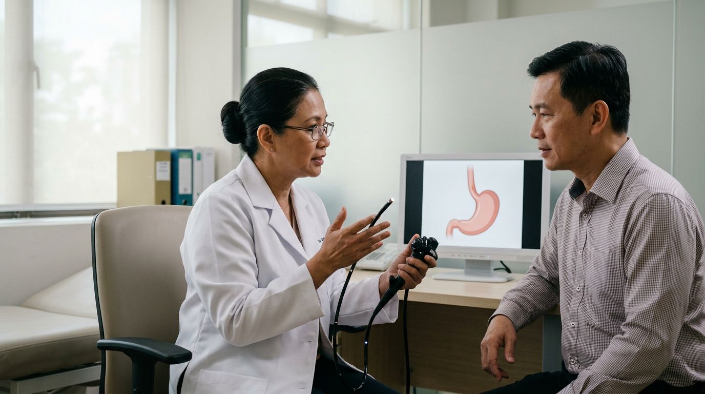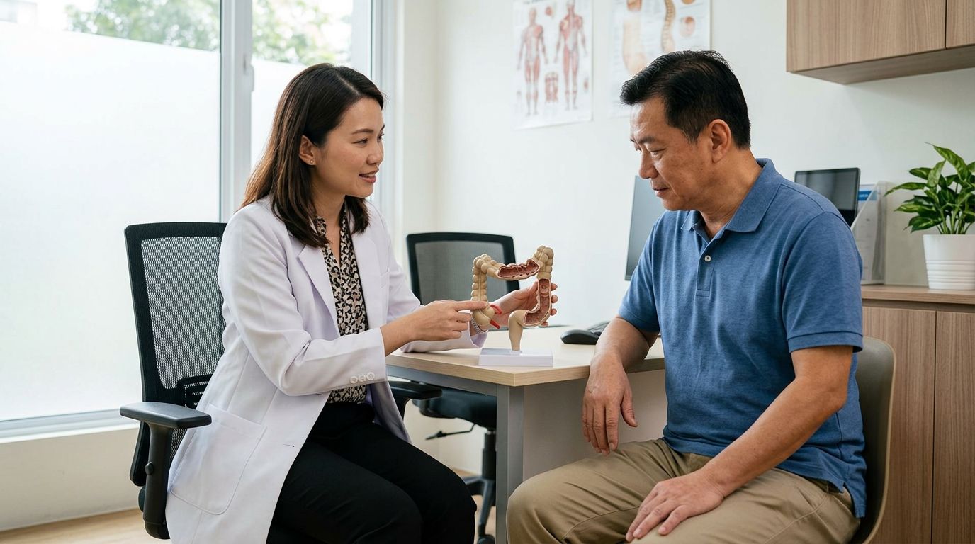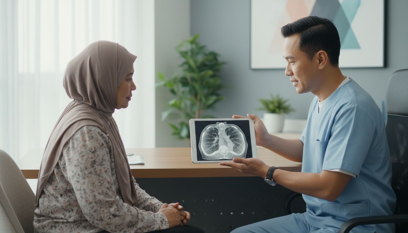
Waiting for scan results can make even a short day feel long. Your lung cancer CT scan results may use unfamiliar words, yet the report is only one part of the clinical picture.
A chest CT scan can show changes in the lungs, lymph nodes, and nearby structures. It cannot confirm cancer on its own. Your doctor uses the scan alongside your symptoms, medical history, blood tests, and sometimes a biopsy to decide what happens next.
Key Takeaways
- A chest CT can detect lung nodules, a mass, enlarged lymph node, fluid, or another change, but these findings do not automatically mean cancer.
- Your radiology report describes what is visible. Your oncologist or respiratory specialist explains what it may mean for you.
- A biopsy for cancer is often needed to confirm a cancer diagnosis and identify the exact cell type.
- Cancer staging may require further imaging, such as a PET-CT scan or MRI scan for cancer, before treatment decisions are made.
- A cancer second opinion or medical report review can help you understand the recommended tests, treatment plan, expected waiting time, and possible costs.
Understanding Lung Cancer CT Scan Results
A computed tomography scan uses X-rays and computer processing to create detailed cross-sectional images. Compared with a standard chest X-ray, CT can show much smaller lung changes and provide a clearer view of their location.
You may have had a low-dose chest CT for low-dose lung cancer screening, a diagnostic CT because of cancer symptoms, or a repeat scan after an earlier finding. The reason for your scan affects how the doctor reads the result.
For example, screening scans are usually performed yearly for people with a higher risk history. A diagnostic scan may use contrast dye and give more information when a doctor is investigating a symptom or an abnormal chest X-ray.
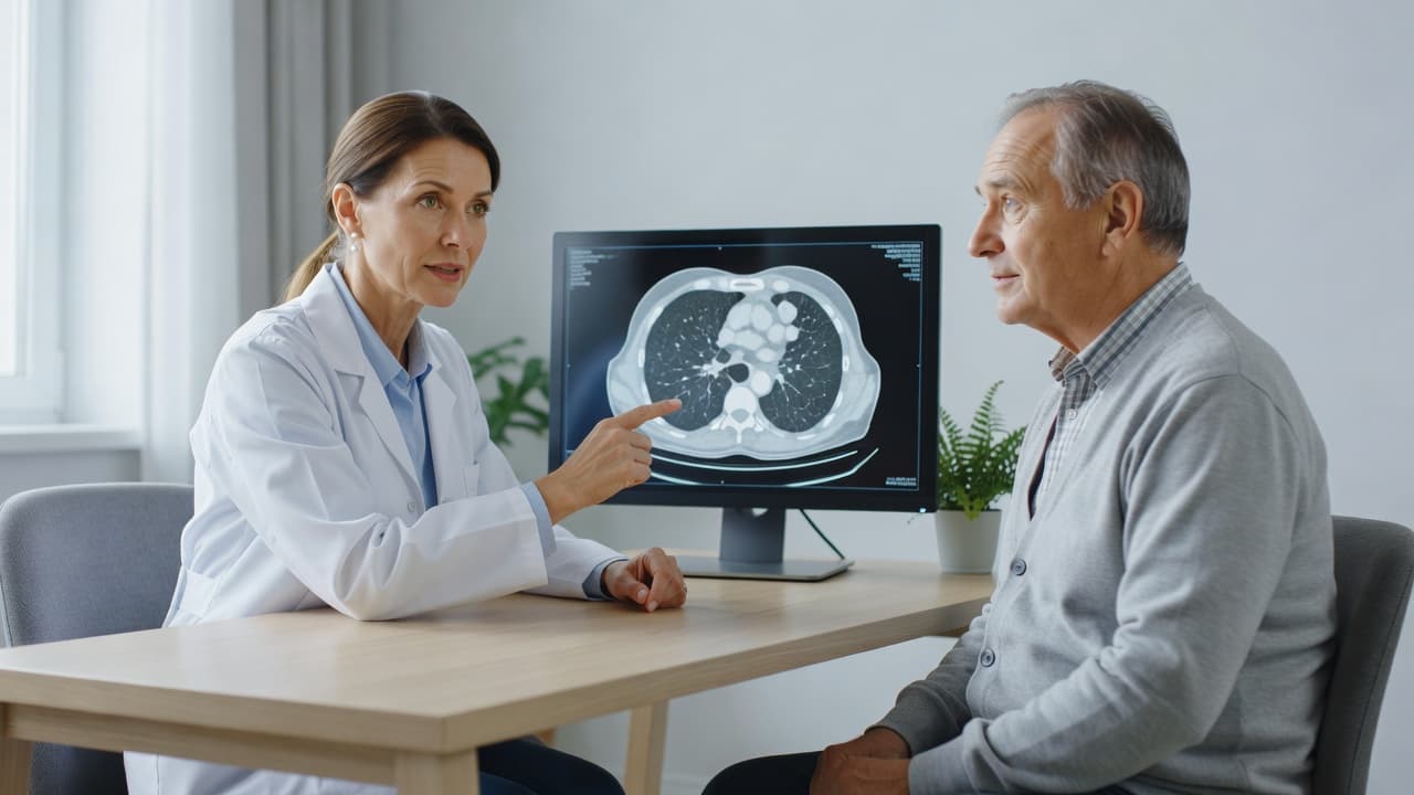
Your report usually has two main sections:
- Findings describe what the radiologist saw, including size, shape, location, and comparison with any older scans.
- Impression gives a short summary of the most important findings and may suggest the next test or follow-up interval.
A scan may show no concerning abnormality. It may also show a small nodule, scar tissue, infection-related inflammation, emphysema, or changes linked to an earlier illness. When evaluating screening exams, a radiologist often applies Lung-RADS as a standardized reporting system to categorize malignancy probability and guide follow-up steps. Some findings are unrelated to the lungs, such as a thyroid nodule or a change in the upper abdomen.
A CT report can identify something that needs attention, but it usually cannot tell you the exact cause without your full clinical information and, in some cases, tissue testing.
If the report says “suspicious for malignancy” or recommends urgent follow-up, contact the doctor who ordered the scan promptly. Try not to interpret one sentence in isolation. Terms such as “cannot exclude” and “recommend correlation” often show that the radiologist needs more information, not that cancer has been confirmed.
Common Words in Lung Cancer CT Scan Results
Radiology language is precise, but it can sound alarming when you read it without explanation. These terms often appear in lung cancer CT scan results.
| Report term | What it usually describes | What may happen next |
|---|---|---|
| Nodule | A small rounded spot in the lung that may be a benign nodule | Comparison with older scans or follow-up CT |
| Mass | A larger area of abnormal tissue | Specialist assessment and possible biopsy |
| Spiculated | Edges that look irregular or star-like | Further imaging or tissue sampling may be advised |
| Ground-glass opacity | A hazy area that does not fully block lung markings | Short-term follow-up or assessment for inflammation |
| Consolidation | Lung tissue filled with fluid, cells, or other material | Treatment for infection or repeat imaging may be considered |
| Enlarged lymph nodes | Lymph nodes larger than expected | PET scan, biopsy, or monitoring depending on context |
| Pleural effusion | Fluid around the lung | Assessment of the cause and possible drainage |
| Stable | No significant change from an earlier scan | Continued surveillance may be recommended |
Size matters, but it is not the only factor. Doctors also evaluate lung nodules by looking at the border, density, growth rate, location, smoking history, age, previous infections, and whether there are enlarged lymph nodes. Radiologists often use standardized Lung-RADS categories to score these imaging findings and guide patient management.
A report may say “interval growth” when a finding has increased compared with an earlier scan. When imaging findings are suspicious for lung cancer, the report will typically recommend specific next steps. Conversely, “stable” means it has not changed enough to meet the radiologist’s threshold for concern. Bring prior scans or reports if they were done at another facility. Comparison can prevent repeat tests and change the recommendation.
The American Lung Association notes that incidental findings can appear during screening and may need separate follow-up. Its guide to what to expect from lung cancer screening explains why an abnormal result does not always point to lung cancer.
When a CT Finding Needs More Tests
A concerning CT scan does not replace a cancer diagnosis. Your doctor may first review older imaging, repeat a CT scan after a defined interval, or refer you to a cancer specialist, respiratory physician, thoracic surgeon, or multidisciplinary cancer hospital team.
When the scan suggests a possible tumour, common next steps in a thorough diagnostic evaluation include:
- PET-CT scan: A PET scan for cancer may show areas with increased metabolic activity and help assess lymph nodes or possible spread. Infection and inflammation can also appear active, so PET results still need clinical interpretation.
- Lung biopsy: A lung biopsy removes cells or tissue for laboratory testing. Depending on the location, this may involve a percutaneous needle biopsy through the chest wall, bronchoscopy, endobronchial ultrasound, or surgery.
- Pathology and biomarkers: A pathology report review identifies the cancer type if cancer is found. Biomarker testing and genetic testing for cancer can look for changes that may affect lung cancer treatment choices.
- Additional scans: Your team may request an MRI scan for cancer, often of the brain, or other confirmatory tests based on the CT findings and your symptoms.
The process can feel slow because each answer shapes the next decision. However, starting treatment without knowing the cell type, stage, and biomarkers can rule out options that may suit you better.
Keep an eye on symptoms while you wait. A persistent or changing cough, coughing up blood, new breathlessness, chest pain, unexplained weight loss, ongoing hoarseness, or repeated chest infections should be reported. These cancer symptoms can have many causes, but new or worsening symptoms deserve timely medical advice.
If you develop severe breathlessness, chest pain, confusion, or cough up a significant amount of blood, seek urgent medical care.
How CT Results Fit Into Cancer Staging
If a biopsy confirms lung cancer, doctors need to know how far it has spread before recommending cancer treatment. This process is called cancer staging.
For non-small-cell lung cancer, teams commonly use the TNM staging system. “T” describes the primary tumor size and local spread. “N” refers to nearby lymph nodes and whether cancer has reached them. “M” indicates whether cancer has spread to distant parts of the body. Small cell lung cancer is often described as limited-stage or extensive-stage, although detailed staging can still guide care.
CT images help map the chest. They can show the tumor’s relationship to airways, blood vessels, chest wall, and lymph nodes. Still, CT cannot always distinguish cancer from inflammation in a lymph node. PET imaging, biopsy, or both may clarify the picture.
Your cancer stages should never be guessed from a CT report alone. The final stage may change after PET imaging, brain MRI, a lymph node biopsy, surgery, or a pathology report review. That is common and does not mean anyone made an error.
Cancer screening scans and scans performed after diagnosis answer different questions. Screening looks for early changes before symptoms develop. A diagnostic work-up looks for the type and extent of a known or suspected cancer. Follow-up scans check whether a treatment plan is working or whether a finding remains stable.
Lung Cancer Treatment After Diagnosis
Your treatment is based on the cancer type, stage, scan findings, biopsy results, pathology report, biomarkers, genetic testing, overall health, lung function, medical history, hospital availability, and your oncologist’s assessment.
For early-stage non-small cell lung cancer, cancer surgery may be considered if you are medically fit for it. A surgeon may remove a small section of lung, a lobe, or sometimes an entire lung. Some centres offer minimally invasive or robotic cancer surgery, but the approach depends on the tumour location and your health.
Radiotherapy, also called radiation therapy, can treat a localised tumour when surgery is unsuitable. Stereotactic body radiotherapy delivers focused treatment over a small number of sessions in selected cases. Proton therapy and MR Linac treatment may be available in certain countries and centres, but availability alone does not determine whether they are appropriate.
As part of effective post-screening management, advanced cancer treatment options for locally advanced or metastatic disease can include chemotherapy, immunotherapy, targeted therapy, or combinations of these. Targeted medicines only work when testing finds a relevant genetic alteration. Immunotherapy suitability depends on several clinical factors, including tumour testing and your health status.
Advanced cancer treatment is not one fixed pathway. A person with a targetable mutation may receive a different first treatment from someone whose pathology shows another subtype. Your oncologist should explain the goal of care, likely side effects, treatment schedule, and how the team will monitor your response.
Reviewing Your Report, Costs, and Hospital Options
Ask for copies of the CT report, scan images, referral letter, pathology report, and blood results. A well-organised folder makes an oncology consultation more productive, especially if your doctor recommends a follow-up observation plan to monitor stable findings over time.
You may want a cancer second opinion when the diagnosis is uncertain, the recommended plan feels unclear, surgery is complex, or you are considering treatment at another centre. A second specialist can review the same evidence and may agree with the first plan. Agreement can bring reassurance, while a different view can lead to useful questions.
When you compare cancer hospitals, ask whether the team includes medical oncology, radiation oncology, thoracic surgery, respiratory medicine, pathology, radiology, palliative care, and specialist nurses. Ask how the team discusses complex cases and who will coordinate your appointments.
Cancer treatment cost varies widely. It can include consultations, biopsy procedures, scans, medicines, surgery, hospital stays, radiotherapy planning, and follow-up. Before deciding where to treat cancer, request a written cancer treatment quotation that separates expected charges from items that may change.
If you are considering overseas cancer treatment, seek coordination support that helps you submit medical reports, understand required documents, and compare practical arrangements. The overseas team must still assess you directly before recommending care. Travel should not delay urgent local treatment without advice from your treating doctor.
Anxiety can also affect sleep while you wait for answers. Gentle routines and practical ways to sleep better may help you rest, but they do not replace medical support for persistent distress.
Questions to Ask at Your Oncology Consultation
Bring someone you trust if possible. They can take notes while you focus on the discussion. These questions can help you understand your next step:
- What does the CT scan show, and which finding is most important?
- Is this finding likely to be cancer, or are there other possible explanations?
- Do I need a repeat CT, PET-CT scan, MRI, bronchoscopy, or lung biopsy?
- If I need a biopsy, which method do you recommend and why?
- Has cancer staging started, and what information is still missing?
- If cancer is confirmed, what type is it and will you order biomarker or genetic testing?
- What lung cancer treatment options fit my situation, and what are their goals?
- How long can I safely wait before the next test or treatment?
- What side effects, practical costs, and travel needs should I expect?
- Would a second opinion change anything at this point?
Write down answers in plain language. If you do not understand a term, ask the doctor to explain it without medical shorthand. You have the right to know why a test or treatment is being suggested.
Final Thoughts
A CT scan can reveal an important clue, but lung cancer CT scan results are not the final diagnosis by themselves. The most useful next step is a timely conversation with the doctor who knows your scans, symptoms, and medical history.
Keep copies of your reports, ask direct questions, and allow the full assessment to guide your choices. Clear information helps you take part in decisions without rushing to conclusions.
Frequently Asked Questions
Does an abnormal lung CT scan mean I have cancer?
No. CT scans can show benign nodules, scars, infection, inflammation, fluid, and other non-cancerous changes. Your doctor may recommend comparison scans, follow-up imaging, PET imaging, or a biopsy depending on the finding.
How long does it take to get CT scan results?
Timing varies by hospital and whether the scan needs urgent review. Some results are available within days, while a full diagnostic work-up can take longer because it may include specialist appointments and additional tests.
Can a CT scan tell the stage of lung cancer?
A CT scan provides important staging information, especially about the lungs and chest lymph nodes. However, doctors may need PET imaging, MRI, biopsy results, or surgical findings before they can confirm the stage.
What does a lung nodule mean?
A lung nodule is a small spot in the lung. Many are harmless, and finding a benign nodule is very common on chest imaging. Your doctor considers its size, appearance, growth, and your personal risk factors before recommending monitoring or further tests when evaluating lung nodules.
Should I get a second opinion after a lung cancer diagnosis?
A second opinion can be helpful before major surgery, when treatment choices are complex, or if you want another specialist to review your scans and pathology. It should support, not delay, time-sensitive care.
Is this article medical advice?
This content is for general information only. It may not be fully verified or apply to every patient, and it is not medical advice, a diagnosis, or a treatment recommendation. You must consult your own doctor, oncologist, or qualified healthcare professional before making any medical decision. SureLah does not take responsibility for medical decisions, treatment outcomes, doctor recommendations, hospital decisions, or actions taken based on this article.