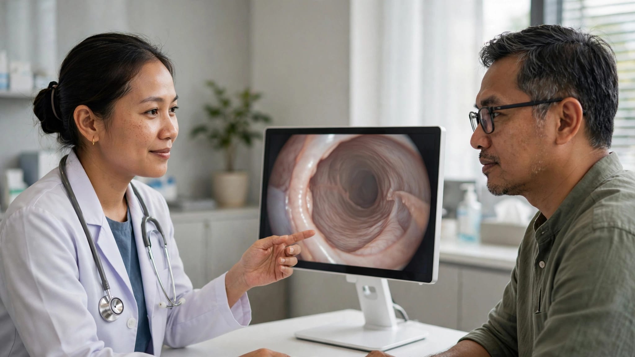
Waiting for oral cancer biopsy results can make every day feel longer. This article can help you prepare for a clear conversation with your doctor, not interpret the report on your own.
Results from oral lesions may be benign, show dysplasia, or confirm cancer in sampled tissue. Timely assessment supports early detection, while an accurate discussion of the report can guide your next step.
Bring a family member if possible, write down your questions, and ask for a copy of the report.
Key Takeaways
- An oral cancer biopsy report describes the tissue that was sampled; it does not independently show the full extent of disease or determine treatment.
- Ask your doctor to explain the exact diagnosis, sample adequacy, dysplasia grade, surgical margins, and any additional tests in everyday language.
- A positive result may lead to staging scans and referral to a multidisciplinary head and neck team, while treatment depends on the tumour and your overall health.
- A benign or inconclusive result does not always explain persistent oral lesions; seek follow-up if symptoms continue, return, or change.
- Keep copies of your pathology report, scans, and treatment plan, and consider a second opinion when the diagnosis or proposed care remains uncertain.
What a biopsy report can tell you
A biopsy allows a pathologist to perform a microscopic examination of cells and tissue from oral lesions. It remains the most reliable way to support early detection, improve diagnostic accuracy, and confirm or exclude oral cancer.
Why the report may take longer than expected
There is no single turnaround time for every oral biopsy. Some reports are ready within several working days, while others take longer when the laboratory needs extra tissue processing, deeper sections, or immunohistochemistry stains.
At a government or private hospital in Malaysia, ask when the report is expected and who will contact you. If additional testing is underway, ask what it is designed to clarify.
Most tissue samples undergo histopathological examination with haematoxylin and eosin, often called H&E staining. In selected cases, immunohistochemistry helps distinguish between cell types or supports the pathological diagnosis.
Terms that deserve a plain-language explanation
A report may describe inflammation, infection, traumatic ulceration, leukoplakia, erythroplakia, epithelial dysplasia, or oral squamous cell carcinoma. Leukoplakia and erythroplakia may be discussed among potential malignant disorders. Appearance alone does not confirm a diagnosis, so you do not need to decode these terms alone.
An incisional biopsy removes a small, representative piece of a lesion. An excisional biopsy removes the entire small lesion where appropriate. Incisional and excisional procedures are commonly performed under local anesthesia when clinically appropriate. A brush biopsy collects cells and can help assess a suspicious area, but an abnormal brush result usually still needs a tissue biopsy for confirmation.
Blood- or saliva-based liquid biopsy approaches are being studied alongside salivary biomarkers and molecular diagnostics. These approaches remain emerging, and their validation and clinical availability vary. A liquid biopsy cannot currently replace a traditional tissue biopsy when oral cancer is suspected. A systematic review of oral biopsy methods describes why the tissue sample remains central to diagnosis.
Questions to ask about your biopsy report
Ask your doctor to explain what the report says about a possible oral cancer diagnosis in everyday language. Prompt clarification supports early detection. Then repeat the key points back in your own words. This helps you leave the consultation knowing what has been confirmed and what remains uncertain.

Confirm the diagnosis and sample
Start by asking what the pathologist found and whether the sample answers the original concern. A tissue biopsy report describes only the sampled tissue. It doesn’t independently establish the disease’s full extent or likely outcome.
Diagnosis
You may want to ask:
- What is the exact diagnosis written in my pathology report?
- If oral cancer is present, is it oral squamous cell carcinoma or another type?
- Does the report describe invasive cancer, and how deeply has it invaded?
- How does the formal wording from the histopathological examination fit with what you saw during my clinical examination?
- What did the microscopic examination of the sample show?
- Were immunohistochemistry tests performed, and what did they add?
- Could a head and neck pathologist review the slides if anything is unclear?
Sample adequacy
- Was this an incisional biopsy or excisional biopsy, was local anesthesia used, and was enough tissue obtained?
- Did the sample adequately represent the oral lesions, and does it answer the original concern?
- Does a liquid biopsy have a validated role in my specific case, or would it only complement rather than replace a tissue diagnosis?
- Is a sentinel lymph node biopsy relevant in my case? It isn’t required for every patient and depends on the tumour and my specialist’s staging plan.
Oral squamous cell carcinoma is the most common cancer type in the oral cavity. A clinical reference on oral squamous cell carcinoma explains the pathology terms your specialist may use.
Ask what dysplasia and surgical margins mean for you
Epithelial dysplasia means abnormal cells are present in the mouth lining. It isn’t the same as invasive cancer, although moderate or severe dysplasia may need closer follow-up because it can carry a higher risk of progression.
Margins matter only when the doctor removes a whole lesion or tumour. They describe the distance between abnormal cells and the edge of the removed tissue. Some reporting systems use more than 5 mm as clear, 1 to 5 mm as close, and less than 1 mm as involved. Your surgical team should explain the definition used in your case.
Ask these questions:
- Does my report show mild, moderate, or severe dysplasia?
- If the lesion was removed, are the margins clear, close, or involved?
- What is the closest margin measurement in millimetres?
- Does the report mention perineural invasion or lymphovascular invasion?
- Does the pathology show carcinoma in situ or invasive disease?
- Do I need another biopsy, excision, or regular monitoring?
Detailed oral-cavity pathology reporting guidance includes many of these features because they can affect follow-up and specialist decisions.
A pathology report names what was found in the sample. Your scans, examination, and lymph node assessment help determine the full diagnosis and next steps.
What happens if the biopsy is positive
A positive biopsy may confirm oral cancer, and that can feel overwhelming. You do not need to decide on treatment during the first conversation. Your doctor should refer you to an appropriate head and neck team, which may include an ENT surgeon, oral and maxillofacial surgeon, oncologist, dentist, speech therapist, and dietitian.

Understand the tests used for cancer staging
Your team may arrange a CT scan, MRI scan, ultrasound of the neck, or a PET scan for cancer when appropriate. These tests help assess the primary tumour, nearby lymph nodes, and possible spread. When disease is found through early detection, the timing and extent of staging may differ.
If a neck node looks suspicious, the team may recommend fine needle aspiration or another biopsy. For selected patients with early disease, the multidisciplinary team may discuss a sentinel lymph node biopsy. The team decides whether it is appropriate.
Ask which tests you need, why each test is recommended, and when the results will be reviewed. A liquid biopsy should not replace imaging, clinical examination, or standard tissue diagnosis. Cancer staging combines findings from microscopic examination of the biopsy with imaging and clinical examination, so it may take time to complete.
Discuss cancer treatment options in context
Once staging is clearer, your team can discuss oral cancer treatment in context. Head and neck cancer treatment may involve cancer surgery, radiotherapy, chemotherapy, targeted therapy, or immunotherapy. The suitable approach depends on tumour location, cancer stage, depth of invasion, lymph nodes, overall health, dental condition, previous treatment, and specialist assessment.
Selected molecular diagnostics may influence specialist decisions in some cases, but they are not routine for everyone. Ask your oncology team what the aim of treatment is and whether treatment should start promptly. Ask how stage, tumour features, and overall health may affect patient prognosis.
You should also ask what support you may need for eating, speech, mouth care, and pain relief. You can request a written outline of the proposed diagnosis and treatment plan.
Before committing, ask what affects your likely cancer treatment cost, including scans, surgery, medicines, dental work, hospital stay, and rehabilitation. A billing or financial counselling team can provide a written estimate based on your recommended care.
For a broad overview of possible pathways to discuss with your doctor, Explore Cancer Treatment Options. It is an informational and coordination resource, not a substitute for your treating specialist’s recommendation.
When a result is benign or inconclusive
A negative result means no oral cancer was identified in the sampled tissue. It doesn’t confirm that persistent oral lesions have resolved or exclude disease elsewhere, especially if the sample didn’t capture the most abnormal area.
Follow up if the mouth lesion remains
Return to your dentist, oral surgeon, or ENT specialist if oral lesions persist, including a sore, red or white patch, lump, unexplained bleeding, or pain. Non-healing sores lasting two to three weeks deserve prompt assessment for early detection, particularly if they are growing or feel firm. A white patch may represent leukoplakia, and a red patch may represent erythroplakia, but appearance alone cannot confirm either condition.
Also seek prompt medical review for a new neck lump, difficulty swallowing, reduced mouth opening, unexplained tooth loosening, or recurrent bleeding. For post-biopsy care, seek urgent medical attention for uncontrolled bleeding, fever, rapidly increasing swelling, or trouble breathing or swallowing.
Ask why the result was non-diagnostic
An inconclusive report may reflect the area sampled, a specimen that was too small or superficial, processing damage, or limitations in the microscopic examination. It doesn’t mean you have cancer, but it may mean your doctor needs more information.
Ask whether your clinical appearance and biopsy result match, since disagreement may affect diagnostic accuracy. If they don’t, ask whether a repeat tissue biopsy, lesion removal, specialist referral, or a short follow-up interval is appropriate. A repeat procedure may be performed with local anesthesia when appropriate. A liquid biopsy cannot by itself resolve a discordant or non-diagnostic tissue result.
When a second opinion may help
An oral cancer second opinion may be reasonable when the diagnosis or proposed care is uncertain, treatment is extensive, or you want greater confidence before a major decision. It may confirm the original plan, clarify the diagnosis, suggest more tests, or identify another medically appropriate approach. If a liquid biopsy is being considered, ask whether it is validated for your situation and would change management.
A second opinion for cancer works best when the next specialist receives the biopsy report, slides if requested, scan reports, images, referral letters, and treatment proposal. Explore Medical Report Review Support if you need help organising records for review coordination.
You may also consider an oncology second opinion if you are deciding where to treat cancer, have questions about treatment availability, or want to understand differences between cancer hospitals. Different centres may have different specialist teams, technologies, costs, waiting times, and support services. Explore Cancer Second Opinion Support for coordination support when seeking another qualified specialist perspective.
Frequently Asked Questions
How long do oral cancer biopsy results take?
Some reports are ready within several working days, while others take longer if the laboratory needs deeper sections, extra tissue processing, or immunohistochemistry. Ask when the report is expected and who will contact you if additional testing is underway.
Does a positive biopsy confirm oral cancer?
A positive tissue biopsy may confirm cancer in the sampled area, often oral squamous cell carcinoma. Your clinical examination, imaging, and lymph node assessment are still needed to determine the full diagnosis and stage.
What does a negative or benign biopsy result mean?
It means that cancer was not identified in the tissue examined, but it does not always explain a persistent lesion or exclude disease elsewhere. Return to your dentist, oral surgeon, or ENT specialist if a sore, patch, lump, bleeding, or pain continues or changes.
Can a liquid biopsy replace a tissue biopsy?
No, a liquid biopsy cannot currently replace a traditional tissue biopsy when oral cancer is suspected. Blood- or saliva-based tests remain emerging, and their availability and validation vary by situation.
When should I seek a second opinion?
A second opinion may help when the diagnosis is unclear, the biopsy result does not match the clinical findings, or treatment is extensive. Ask whether the specialist can review your pathology report and slides, scans, and proposed treatment plan.
A clear next step
Oral cancer biopsy results are a starting point for informed decisions, not a full treatment plan on their own. Ask for the exact diagnosis and whether the sample was adequate. Ask what unfamiliar report terms mean, what the microscopic examination shows, and which follow-up tests or referrals come next.
If symptoms persist or new changes develop, seek review promptly. Early detection can matter when mouth changes continue or return.
A liquid biopsy may offer additional information as testing evolves, but it doesn’t replace the report or standard diagnostic process.
Keep copies of every report and scan result. Clear records make follow-up appointments, treatment discussions, and a possible second opinion easier to manage.
Medical disclaimer: This article is for general educational information only and may not apply to every patient. It is not medical advice, a diagnosis, or a treatment recommendation. Consult your own doctor, oncologist, or qualified healthcare professional before making medical decisions.